AI-Powered Intraoral Camera Analysis for Pets: How Vets Catch Dental Disease Earlier
Learn how AI-assisted intraoral cameras help veterinarians spot gingivitis, tartar, and early periodontal disease in dogs and cats—improving triage, documentation, and follow-up.
AI-Powered Intraoral Camera Analysis for Pets: How Vets Catch Dental Disease Earlier
Dental disease is one of the most common (and most overlooked) health issues in dogs and cats. An AI intraoral camera analysis for pets workflow—where clear mouth photos or short video clips are captured during a visit and analyzed by computer vision—can help clinics document problems earlier, communicate findings better, and prioritize which patients need a full dental workup.
This article explains how AI-assisted intraoral imaging fits into veterinary dentistry, what it can (and can’t) detect, and how it complements tools like dental radiographs and dedicated scanners.
Why intraoral imaging matters in everyday pet dentistry
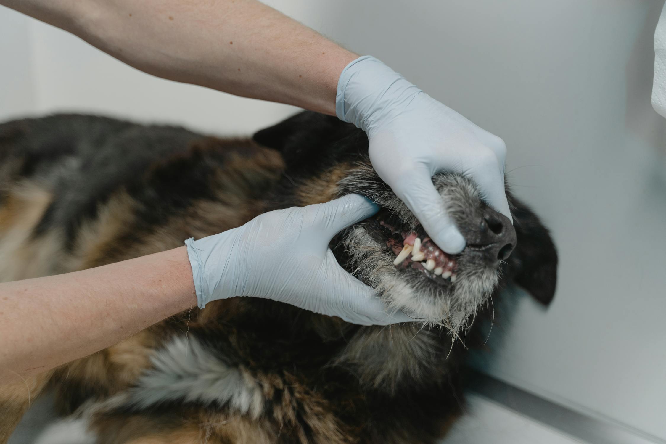
Intraoral cameras are small, maneuverable cameras designed to capture detailed images inside the mouth: incisors, canines, premolars, molars, gumlines, and visible calculus. In routine appointments, they help with:
- Better documentation: baseline images make it easier to track progression over time.
- Owner communication: a clear photo of inflamed gingiva is more convincing than a verbal description.
- Triage: identifying obvious plaque, fractures, or severe gingivitis can help decide next steps.
However, photos alone can be inconsistent—lighting, motion blur, saliva, and angles vary. That’s where veterinary dental imaging AI can add value.
What AI can detect from intraoral photos (and what it can’t)
Computer vision models trained on labeled veterinary dental images can be used to flag patterns associated with common findings, such as:
- Visible tartar/calculus distribution (especially along the gingival margin)
- Gingival inflammation patterns (redness, swelling, bleeding risk)
- Crowding / malocclusion indicators (helpful for documenting)
- Obvious tooth fractures or missing teeth (where visible)
- Halitosis risk markers (indirect signals like heavy calculus)
That said, there are hard limits:
- AI cannot “see” below the gumline from a photo. Periodontal pockets, root pathology, and many resorptive lesions require probing and/or radiographs.
- Cats and tooth resorption: many resorptive lesions are not reliably diagnosed without dental radiographs.
- AI outputs are typically decision support, not a diagnosis.
A practical mindset: use AI to improve consistency and earlier identification of “needs a dental” patients, then confirm with standard veterinary dental protocols.
How AI-assisted intraoral camera analysis supports early periodontal disease detection
When used consistently (e.g., at wellness visits or pre-anesthetic evaluations), AI-assisted imaging can boost early detection periodontal disease pets workflows by:
- Standardizing image capture (recommended angles for each quadrant)
- Highlighting areas of concern (heatmaps/markers on gumline)
- Generating structured notes (e.g., “heavy calculus on maxillary premolars”)
- Tracking change over time (progression score compared to last visit)
This is especially useful for busy clinics where subtle gingivitis can be easy to miss, or where dental recommendations are under-accepted by owners.
A simple clinic workflow (capture → analyze → explain → plan)
A lightweight workflow can look like this:
- Capture: technician takes 6–10 quick intraoral shots (left/right, upper/lower, front teeth).
- Analyze: AI model returns a preliminary assessment (e.g., calculus score, inflammation score, flagged teeth).
- Explain: veterinarian reviews images with the owner using a chairside screen.
- Plan: decide whether to schedule a dental cleaning, recommend home care, or proceed to radiographs.
If your practice already uses AI support for imaging, this can integrate naturally with broader tools like scanners and diagnostic systems. For background, see our overview of diagnostic performance: AI Pet Dental Diagnosis Accuracy.
How this compares to dental radiographs and dedicated dental scanners
Intraoral camera + AI is best thought of as a front-line documentation and triage tool.
- Dental radiographs remain the gold standard for evaluating roots, bone loss, and resorptive disease. AI can sometimes help interpret radiographs, but images are still required. If you’re weighing approaches, compare: AI vs Traditional Dental Exams.
- Dedicated dental scanners (for structured capture and analysis) can offer more standardized datasets than ad-hoc photos. See: AI Pet Dental Health Scanner: How It Works.
A clinic might start with intraoral camera analysis for better documentation, then expand into radiograph workflows and advanced scanning as needed.
Quality, safety, and compliance considerations
To make AI outputs reliable, clinics should pay attention to:
- Image quality: consistent lighting, focus, and minimal glare.
- Annotation and review: final assessments should be clinician-reviewed.
- Data privacy: avoid storing identifying client data in image filenames; ensure vendors follow appropriate security practices.
For authoritative veterinary dentistry standards and training resources, the American Veterinary Dental College is a useful reference: https://avdc.org/
Practical tips for pet owners (what you can do at home)
Even with better clinic tools, prevention matters:
- Brush with pet-safe toothpaste (start slowly and build a routine).
- Use vet-approved dental diets or chews when appropriate.
- Watch for early signs: bad breath, red gums, pawing at the mouth, drooling, or dropping food.
If you want a baseline home-care checklist, see: Dog Dental Care Home Guide.
Conclusion: better visuals lead to earlier care
AI-assisted intraoral imaging won’t replace a full dental evaluation, but it can dramatically improve how early problems are spotted, documented, and explained. By pairing consistent intraoral photos with veterinary dental imaging AI, clinics can increase dental acceptance, reduce missed disease, and guide owners toward timely treatment.
If you’re exploring a more standardized approach to pet oral health screening and documentation, Nerovet is building tools designed for modern veterinary workflows.
Frequently Asked Questions
How accurate is AI dental analysis for pets?
AI dental analysis systems like Nerovet achieve high accuracy rates in detecting common dental conditions such as periodontal disease, tooth fractures, and resorptive lesions. The AI serves as a decision-support tool that helps veterinarians identify issues they might otherwise miss, rather than replacing professional judgment.
How does AI pet dental scanning work?
AI pet dental scanning uses deep learning algorithms trained on thousands of veterinary dental images. When you upload a dental X-ray or intraoral photo, the AI analyzes it to detect anomalies, classify conditions, and generate a detailed report with findings and recommendations.
Can AI replace a veterinary dentist?
No, AI is designed to assist veterinarians, not replace them. AI excels at pattern recognition and can flag potential issues for review, but clinical decision-making, treatment planning, and procedures still require a trained veterinary professional.
Want to apply this workflow in your clinic?
Book a Nerovet demo to see practical workflow recommendations for dog and cat dental imaging.