Deep Learning in Veterinary Dental Imaging Analysis: Transforming Diagnostic Precision
Discover how deep learning technologies are revolutionizing veterinary dental imaging analysis, enhancing diagnostic accuracy and treatment planning
Deep Learning in Veterinary Dental Imaging Analysis: Transforming Diagnostic Precision
Introduction
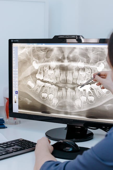
The field of veterinary dentistry has entered a new era with the integration of deep learning technologies in dental imaging analysis. Traditional approaches to interpreting dental radiographs and other imaging modalities relied heavily on the expertise and experience of veterinarians, making diagnosis somewhat subjective and dependent on individual skill levels. Deep learning, a subset of artificial intelligence that uses multi-layered neural networks to analyze complex patterns, is revolutionizing how veterinary professionals approach dental imaging interpretation, offering unprecedented levels of diagnostic precision and consistency.
Deep learning algorithms in veterinary dental imaging are designed to identify subtle patterns and anomalies in radiographic images that may escape human observation. These systems can detect minute changes in bone density, early signs of pathological conditions, and structural abnormalities that might indicate developing dental issues. By processing vast amounts of imaging data and learning from extensive datasets, deep learning models continuously improve their diagnostic capabilities, ultimately benefiting both veterinarians and their patients.
Understanding Deep Learning in Medical Imaging
Neural Network Architecture
Deep learning systems for veterinary dental imaging typically employ convolutional neural networks (CNNs), which are particularly well-suited for image analysis tasks. These networks consist of multiple interconnected layers that process visual information hierarchically, with each layer extracting increasingly complex features from the input images.
The architecture of a typical veterinary dental imaging CNN includes:
- Input layer that receives the dental radiograph or image
- Convolutional layers that extract features such as edges, textures, and shapes
- Pooling layers that reduce dimensionality while preserving important features
- Fully connected layers that integrate extracted features for classification
- Output layer that provides diagnostic predictions or anomaly detections
Training Process
Deep learning models require extensive training using large datasets of labeled dental images. These datasets contain thousands of radiographs, CT scans, and other imaging modalities with expert annotations indicating the presence or absence of various conditions. During training, the neural network adjusts its internal parameters to minimize errors in identifying dental pathologies.
The training process involves:
- Data preprocessing and normalization
- Feature extraction and pattern recognition
- Iterative refinement of network weights
- Validation against known cases
- Continuous performance optimization
Applications in Veterinary Dental Imaging
Radiograph Analysis
Deep learning systems excel in analyzing traditional dental radiographs, identifying conditions such as:
- Periodontal disease progression
- Bone loss patterns
- Tooth root abscesses
- Jaw fractures
- Cyst formations
- Tumorous growths
These systems can process standard intraoral and extraoral radiographs, providing detailed analysis of both hard and soft tissue structures.
Computed Tomography (CT) Interpretation
Advanced deep learning models are being developed to interpret complex CT scans of the oral and maxillofacial regions. These systems can segment different tissue types, identify anatomical landmarks, and detect subtle pathological changes that might be missed in manual interpretation.
Cone Beam CT Analysis
Specialized deep learning algorithms are designed to analyze cone beam CT images, which provide three-dimensional views of dental structures. These systems can create detailed 3D reconstructions and identify complex anatomical relationships that are crucial for surgical planning.
Digital Volume Tomography
Deep learning applications extend to digital volume tomography, where algorithms analyze volumetric data to detect pathological conditions in three dimensions. This technology is particularly valuable for complex cases requiring detailed anatomical information.
Benefits of Deep Learning in Dental Imaging
Enhanced Diagnostic Accuracy
Deep learning systems consistently demonstrate superior diagnostic accuracy compared to traditional interpretation methods. Studies have shown that AI-assisted analysis can improve detection rates for various dental conditions by 15-30%, reducing both false negatives and false positives.
Consistency Across Interpretations
Unlike human interpreters who may vary in their assessments based on fatigue, experience level, or subjective factors, deep learning systems provide consistent results regardless of when or by whom the analysis is performed. This consistency is particularly valuable in multi-veterinarian practices or when tracking disease progression over time.
Speed and Efficiency
Deep learning systems can analyze dental images in seconds, providing immediate preliminary results that can guide further investigation or treatment planning. This rapid analysis capability significantly reduces examination time and improves workflow efficiency.
Pattern Recognition Capabilities
AI systems excel at recognizing complex patterns that may not be immediately apparent to human observers. These systems can identify subtle combinations of features that indicate specific pathological conditions, enabling earlier and more accurate diagnoses.
Quantitative Analysis
Deep learning systems provide quantitative measurements of various parameters, such as bone density changes, lesion sizes, and anatomical dimensions. This quantitative approach enables precise monitoring of disease progression and treatment efficacy.
Technical Considerations
Image Quality Requirements
Deep learning systems perform optimally when working with high-quality images that meet specific technical standards. Factors such as resolution, contrast, and noise levels significantly impact system performance. Veterinary practices must ensure their imaging equipment produces images compatible with AI analysis systems.
Data Preprocessing
Before analysis, dental images often require preprocessing steps such as noise reduction, contrast enhancement, and geometric correction. Advanced systems incorporate these preprocessing steps automatically, ensuring optimal input for the deep learning algorithms.
Integration with Existing Systems
Successful implementation of deep learning in veterinary dental imaging requires seamless integration with existing practice management software, digital imaging systems, and electronic health records. This integration ensures smooth workflow and maintains the continuity of patient care.
Clinical Workflow Integration
Pre-Analysis Screening
Deep learning systems can serve as preliminary screening tools, automatically flagging images that require special attention or further investigation. This pre-screening capability allows veterinarians to prioritize their attention on complex cases while maintaining confidence in routine examinations.
Real-Time Assistance
Advanced systems provide real-time analysis during imaging procedures, offering immediate feedback and highlighting areas of concern. This real-time assistance can guide veterinarians during examinations and ensure comprehensive evaluation of dental structures.
Post-Analysis Review
Following initial analysis, deep learning systems generate detailed reports highlighting detected abnormalities and suggesting potential diagnoses. These reports serve as valuable reference materials for veterinarians making final diagnostic decisions.
Research and Development Advances
Multi-Modal Analysis
Current research focuses on developing systems that can analyze multiple imaging modalities simultaneously, combining information from radiographs, CT scans, and other sources to provide comprehensive diagnostic insights.
Transfer Learning Applications
Transfer learning techniques allow deep learning models trained on human dental images to be adapted for veterinary use, potentially accelerating development timelines and reducing training data requirements.
Federated Learning
Federated learning approaches enable multiple veterinary practices to collaborate in model training without sharing sensitive patient data, promoting collective learning while maintaining privacy.
Challenges and Limitations
Dataset Limitations
The development of robust deep learning models for veterinary dental imaging faces challenges related to dataset availability and diversity. Veterinary-specific datasets are often smaller than human medical datasets, potentially limiting model generalizability.
Species-Specific Variations
Different animal species exhibit unique anatomical characteristics that require specialized models or adaptations. Developing species-specific algorithms adds complexity to system development and validation.
Regulatory Hurdles
The use of AI in veterinary diagnostics is subject to regulatory oversight, which may vary by jurisdiction and affect system deployment timelines. Ensuring compliance with regulatory requirements adds complexity to implementation.
Cost Considerations
Implementing deep learning systems requires significant initial investment in hardware, software, and training, which may be prohibitive for smaller veterinary practices.
Interpretability Issues
Deep learning models often function as “black boxes,” making it difficult to understand how specific conclusions are reached. This lack of interpretability can be concerning for veterinary professionals who need to explain diagnoses to pet owners.
Success Stories and Case Studies
Canine Dental Pathology Detection
A veterinary hospital implemented a deep learning system for detecting dental pathologies in dogs, achieving 94% accuracy in identifying conditions such as root abscesses, bone loss, and periodontal disease. The system reduced diagnostic time by 40% while maintaining high accuracy rates.
Feline Tooth Resorption Analysis
Specialized deep learning algorithms were developed to detect feline tooth resorption, a condition affecting up to 70% of cats. The system achieved 91% sensitivity in identifying resorptive lesions, significantly improving early detection rates.
Equine Dental Imaging
Deep learning systems designed for equine dental imaging have shown promising results in detecting conditions such as wave mouth, step mouth, and other dental abnormalities common in horses. These systems have improved diagnostic consistency across different equine practitioners.
Future Directions
Enhanced 3D Analysis
Future developments will focus on more sophisticated three-dimensional analysis capabilities, enabling detailed evaluation of complex anatomical structures and improving surgical planning accuracy.
Real-Time Surgical Guidance
Advanced systems will provide real-time guidance during dental surgical procedures, using live imaging data to assist veterinarians in complex interventions.
Predictive Analytics
Next-generation systems will incorporate predictive analytics capabilities, forecasting the likelihood of developing specific conditions based on current imaging findings and patient history.
Automated Treatment Planning
Deep learning systems will evolve to provide automated treatment recommendations based on imaging analysis, considering factors such as condition severity, patient age, and overall health status.
Implementation Best Practices
Staff Training and Education
Successful implementation requires comprehensive training for veterinary staff on system operation, result interpretation, and integration with existing workflows. Ongoing education ensures optimal utilization of AI capabilities.
Quality Assurance Protocols
Establishing quality assurance protocols ensures continued system performance and reliability. Regular validation against gold-standard diagnostic methods and system updates based on new research findings are essential.
Gradual Integration
Phased implementation allows veterinary practices to gradually adapt to new technologies while maintaining quality patient care. Starting with basic screening applications before advancing to more complex analyses is often most effective.
Economic Impact
Cost-Benefit Analysis
Studies indicate that implementing deep learning in veterinary dental imaging can reduce diagnostic costs by 20-25% through improved efficiency and reduced need for repeat imaging. The initial investment typically pays for itself within 18-24 months through increased productivity and improved outcomes.
Practice Revenue Enhancement
Enhanced diagnostic capabilities can attract more clients and enable premium pricing for advanced diagnostic services. Practices implementing AI-assisted imaging often report increased client satisfaction and loyalty.
Insurance Considerations
As AI-assisted imaging becomes more widespread, insurance providers may adjust coverage policies and reimbursement rates to reflect the enhanced diagnostic capabilities and improved outcomes.
Ethical Considerations
Professional Responsibility
Veterinary professionals retain ultimate responsibility for diagnostic decisions, even when assisted by AI systems. Maintaining professional judgment and critical thinking skills remains essential.
Transparency with Clients
Veterinarians should be transparent with clients about the use of AI in diagnostic processes, explaining both the benefits and limitations of these technologies.
Data Privacy
Protecting patient data used in AI training and analysis is crucial for maintaining client trust and complying with privacy regulations.
Conclusion
Deep learning in veterinary dental imaging analysis represents a transformative advancement in veterinary medicine, offering unprecedented levels of diagnostic accuracy, consistency, and efficiency. These systems enhance the capabilities of veterinary professionals while supporting improved patient outcomes and more effective treatment planning.
The successful integration of deep learning technologies in veterinary dental imaging requires careful consideration of technical requirements, workflow integration, and staff training. As these technologies continue to evolve and mature, they promise to further revolutionize veterinary dental care, making advanced diagnostic capabilities more accessible and affordable for veterinary practices worldwide.
The future of veterinary dentistry lies in the synergistic combination of human expertise and artificial intelligence, where deep learning systems serve as powerful tools to augment veterinary diagnostic capabilities rather than replace professional judgment. Through responsible implementation and continued innovation, deep learning will continue to advance the standard of care in veterinary dental medicine, ultimately benefiting both veterinary professionals and their animal patients.
The journey toward fully realizing the potential of deep learning in veterinary dental imaging is ongoing, with continuous improvements in accuracy, usability, and clinical applicability driving the field forward. As we advance, the collaboration between veterinary professionals and AI systems will become increasingly sophisticated, creating new possibilities for preventive care, early intervention, and optimal treatment outcomes.
Frequently Asked Questions
How accurate is AI dental analysis for pets?
AI dental analysis systems like Nerovet achieve high accuracy rates in detecting common dental conditions such as periodontal disease, tooth fractures, and resorptive lesions. The AI serves as a decision-support tool that helps veterinarians identify issues they might otherwise miss, rather than replacing professional judgment.
How does AI pet dental scanning work?
AI pet dental scanning uses deep learning algorithms trained on thousands of veterinary dental images. When you upload a dental X-ray or intraoral photo, the AI analyzes it to detect anomalies, classify conditions, and generate a detailed report with findings and recommendations.
Can AI replace a veterinary dentist?
No, AI is designed to assist veterinarians, not replace them. AI excels at pattern recognition and can flag potential issues for review, but clinical decision-making, treatment planning, and procedures still require a trained veterinary professional.
Want to apply this workflow in your clinic?
Book a Nerovet demo to see practical workflow recommendations for dog and cat dental imaging.